(Poster 23) Pelvic Organ Prolapse During Pregnancy a Giant Bladder Stone. Case Report
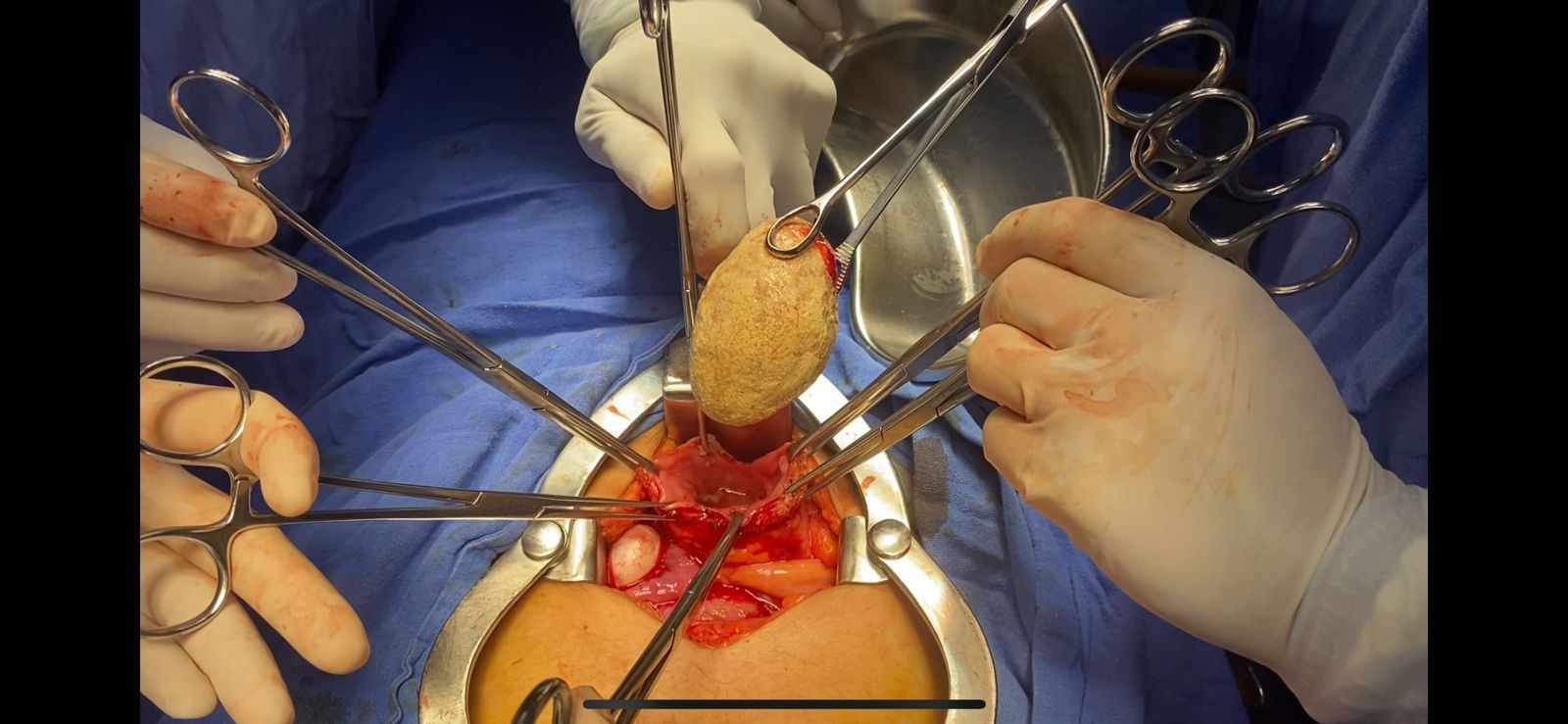
cystolithotomy
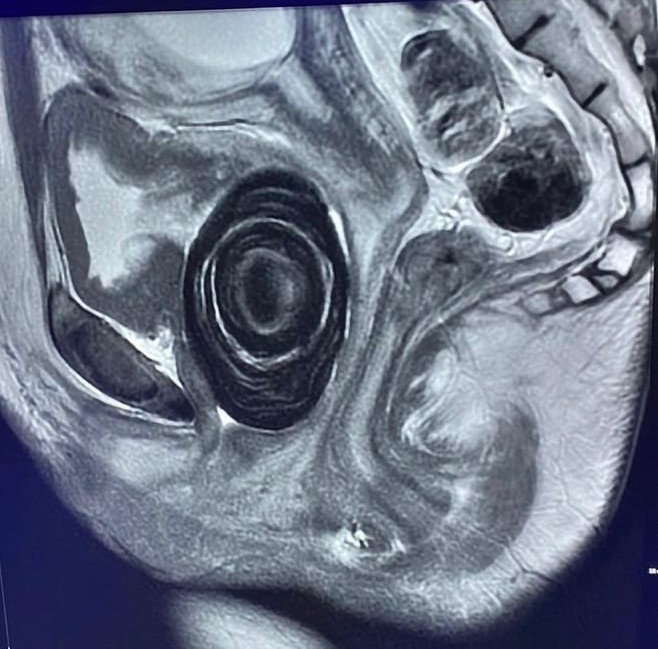
PELVIC MR / BLADDER STONE
Objectives: A 19-year-old primiparous woman with a 13 week pregnancy without prenatal care presents with a sensation of a foreign body in her vagina that has been developing for 7 years, gradually growing, and the need to finger to urinate and intercourse . She begins with dysuria, foul-smelling vaginal discharge, and intense cramping pain. Examination reveals a stony tumor in the anterior vaginal wall measuring approximately 5x6 cm POP IIBA classification, but the transurethral catheter fails to pass, with purulent discharge upon acupressure.
An ultrasound examination revealed a bladder with concentric mural thickening of 20 mm, adjacent to the bladder floor. An oval lesion with calcified edges and a posterior acoustic shadow measuring 4 x 3.7 cm was identified. Left kidney stones associated with grade III hydronephrosis, and a live fetus of 13.5 weeks . MRI identified a 3.9 x 3.9 x 6.6 cm oval lesion with a laminated appearance, impacted on the external urethral sphincter.
Methods: An open cystolithotomy was performed, removing a 7x6 cm stone weighing 160 grams, which was impacted on the bladder floor. A transurethral catheter and diversion cystostomy were placed, continuing the antibiotic regimen for 7 days with a carbapenem. The catheters were maintained for 3 weeks without any obstetric complications.
Results: Stones in pregnancy represent less than 0.5% of cases and are associated with voiding obstruction, recurrent urinary tract infections, metabolic disorders, malnutrition, and male sex (1). Giant bladder stones are those weighing more than 100 grams, and stones of this size are often rare findings in contemporary practice that require surgical management (2) (3). In the previous case, the patient presented with recurrent urinary tract infections and malnutrition, which were her main risk factors, leading to obstructive and infectious complications and preterm delivery. The diagnostic approach should include metabolic evaluation to identify the probable etiology and establish measures to prevent recurrence (4).
Conclusions: Giant bladder stones are a rare condition that must be diagnosed and treated promptly to prevent complications associated with obstructive and infectious processes. They require a multidisciplinary approach. In the case of large stones, suprapubic cystotomy should be considered the treatment of choice.